By: Michael Kelberman
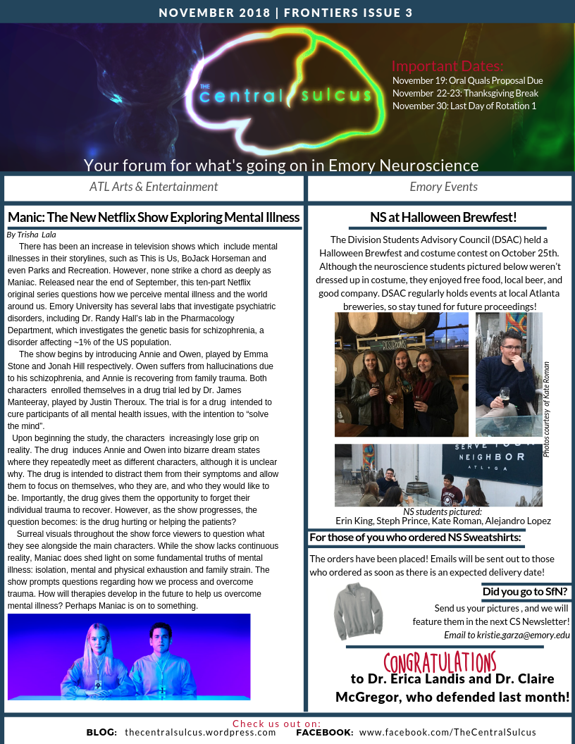
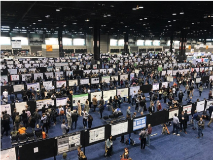
This year, the Emory Neuroscience Community had a large representation at the Annual Society for Neuroscience meeting in Chicago. These included talks at nanosymposiums and a host of poster presentations. With over 25 thousand attendees this year, there is a lot of science that students and faculty presented and a lot more that people may have missed. Here, we will focus on some of the graduate student’s favorite posters at the annual meeting.
Name: Michael Kelberman
Year: 2nd
Mentor: David Weinshenker
Poster Title: Axolotl Homologs for Human Neurodegenerative Proteins
My dissertation work in Dr. Weinshenker’s lab is focused on dysfunction of the locus coeruleus norepinephrine system in the context of Alzheimer’s disease (AD). In the past 15 years, no new drugs have been approved to curb the symptoms of AD. Part of the reason is that AD is a very complex disease and incorporating all aspects of disease into a single model has remained elusive. Thus, development of new models is required to study different aspects of the disease. One condition that has been particularly elusive is tau aggregation. Tau becomes hyperphosphorylated and deposits as neurofibrillary tangles in AD and is consistently reported as a better predictor of cognitive decline and cell death compared to amyloid pathology. Moreover, tau aggregation is associated with other neurodegenerative diseases like Pick’s disease and progressive supranuclear palsy.
At this year’s Society for Neuroscience Conference in Chicago, I attended a poster entitled “Axolotl Homologs for Human Neurodegenerative Proteins”. The presenter, Lucas James, identified a transcript homolog of human tau in the axolotl genome. Although the N-terminal domain was quite different, the binding domain was 94% conserved across species. Moreover, the proline ratios in highly enriched regions were also similar between axolotl and human tau. Axolotls are primarily used in research to study development and regeneration, owing to their remarkable ability to regrow limbs and large portions of their brains. However, these results suggest they may also be useful in studying AD and other tau-associated diseases.

Name: Liz Heaton
Year: 2nd
Mentor: Shannon Gourley
Poster Title: Selective deletion of the melanocortin-4 receptor in the prefrontal cortex alters feeding and executive function-like behavior
My dissertation work is primarily focused on the role of the melanocortin-4 receptor (MC4R) in familiar, habitual behaviors. MC4R is classically studied in the hypothalamus where it is important for regulating satiety signals. However, new research in the past ten years has shown that MC4R expression in the brain is widespread, and MC4R function can differ by brain region. My lab recently compared gene expression in the dorsomedial striatum (DMS) between mice that could and could not break established habits, revealing that lower levels of MC4R are associated with the ability to break habits. These experiments piqued my interest in the function of MC4R both within the DMS and in regions that project to the DMS, such as the prefrontal cortex.
At this year’s Society for Neuroscience conference in Chicago, I attended a poster out of Dr. Rachel Ross’s lab at Beth Israel Deaconess on the function of MC4R in the medial prefrontal cortex (mPFC). They found that knocking down Mc4r in the mPFC decreases the excitability of mPFC pyramidal cells, resulting in increased anxiety-like behavior and decreased fear extinction. Prefrontal MC4R-expressing cells were primarily expressed in the prelimbic cortex, a region that sends profuse projections to the DMS. This study highlights one of the novel functions of MC4R outside of the hypothalamus and broadens our understanding of what this receptor actually does in the brain.
Name: Thomas Shiu
Year: 3rd
Mentor: Andrew Escayg
Poster Title: AAV engineering by multiplexed-create selection and rational peptide insertion yields variants with enriched targeting of CNS astrocytes upon systemic delivery
While my research interest is epilepsy, SFN provides an opportunity for me to explore other cutting-edge research in different areas of neuroscience.
My favorite poster at SFN this year came from Xinhong Chen, a graduate student in the Gradinaru group at Cal Tech. The poster presenter discussed an efficient recombinant Adeno-associated virus (rAAV) that can preferentially target astrocytes through systemic intravenous injection. rAAVs are the most commonly used vectors for gene transfer in the nervous system, enabling delivery of fluorescent proteins to mark specific cell types, opsins or DREADDs for neuromodulation, and calcium or voltage indicators for imaging. Furthermore, since rAAV injection has minimal side effects, rAAVs hold great translational value for gene therapy. Since rAAVs pass through the blood brain barrier (BBB) with low efficiency, and to limit and potential off-target effects, these vectors are predominantly delivered through site-directed intracranial injections. However, local injections are insufficient to target larger neurocircuits and/or make it difficult to test gene therapies in diseases that involve the entire nervous system, such as Huntington’s disease.
Gradinaru’s group previously developed a strategy, called Cre-recombination-based AAV targeted evolution (CREATE), to develop rAAVs that can pass through the BBB (Chen et al. Nat Neuro 2017). This method creates a platform for selecting and screening the optimal capsids that can efficiently pass through the BBB without targeting other tissues or systems (such as liver). In this poster, they optimized the CREATE platform and engineered a newer version of an AAV capsid that can target specifically astrocytes in the CNS. While they performed their initial experiment using C57BL/6J mice, they also demonstrated strain-dependent differences in vector efficiency. For instance, the designed rAAV has lower efficiency in BALB/c mice compared to C57BL/6J. This result will inform us to choose more suitable genetic backgrounds for experiments. Chen is planning to deposit the plasmid at Addgene, providing an additional toolbox to target astrocytes noninvasive to the neuroscience community.
Name: Kate Heffernan
Year: 2nd
Mentor: Adriana Galvan, Yoland Smith
Poster Title: Cell-type specific optical recordings of electrophysiological oscillations in behaving mice using genetically encoded fluorescent voltage indicators
My work in Adriana Galvan and Yoland Smith’s labs is focused on astrocyte-neuron interactions in the basal ganglia. The lab employs electrophysiological, optogenetic, chemogenetic, and anatomical methods to understand neuronal network dysfunction in Parkinson’s disease. I am always on the lookout for new techniques that can help us understand basal ganglia functions and anatomy.
Our lab is interested in field potential dynamics, as they can be indicative of different brain states, both in health and disease. Field potentials generally reflect the activity of multiple cell types and current experimental methods are unable to dissect contributions of specific cell types. At this year’s Society for Neuroscience Conference in Chicago, I attended a poster presentation addressing this problem. The poster was entitled “Cell-type specific optical recordings of electrophysiological oscillations in behaving mice using genetically encoded fluorescent voltage indicators”. Dr. Mark Schnitzer’s lab developed a technique termed trans-membrane electrical measurements performed optically (TEMPO) which is used in freely behaving animals. The technique allows for simultaneous measurement of network trans-membrane voltage dynamics in a specific neuronal population and background noise from hemodynamics and motion artifacts. Subsequent analyses allow for the isolation of the voltage-dependent signals from physiological artifacts, making TEMPO significantly more sensitive than other methods. TEMPO can be applied in normal and pathological conditions to ascertain the contributions of specific cell types to different brain states.